
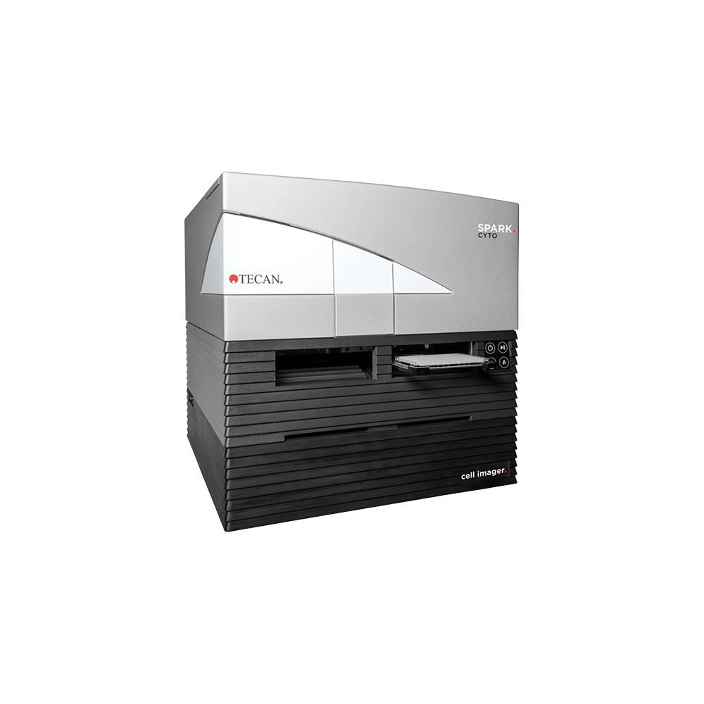
Spark® Cyto
The Tecan Spark® Cyto is a multimode microplate reader combining advanced fluorescence imaging, real-time cell cytometry and established detection technologies in one versatile platform. Designed for 2D and 3D cell-based research, it enables researchers to monitor cell populations, assess cellular responses and collect quantitative and qualitative data within the same instrument. Its imaging system features three magnification levels, four fluorescence channels and brightfield imaging, with whole-well imaging of 96- and 384-well plates using a single image without tiling. Integrated environmental controls, including temperature regulation, CO₂ and O₂ control, and humidity management, support long-term live-cell experiments. Controlled through SparkControl™ software and supported by Image Analyzer™ tools, Spark Cyto offers automated image acquisition and analysis for applications ranging from cell viability and apoptosis to spheroid and organoid research.

Features
- Combines multimode plate reading, fluorescence imaging and real-time cell cytometry.
- Supports both 2D and 3D cell-based research applications.
- Three imaging objectives: 2x, 4x and 10x magnification.
- Four fluorescence channels: blue, green, red and far red.
- Brightfield and digital phase contrast imaging.
- Whole-well imaging of 96- and 384-well plates using a single image.
- Integrated LED-based autofocus system.
- Temperature control up to 42°C.
- Dynamic CO₂ and O₂ control for live-cell experiments.
- Humidity control with integrated Humidity Cassette and Lid Lifter™.
- Multiple detection modes, including absorbance, fluorescence, luminescence, FRET, TR-FRET and fluorescence polarisation.
- SparkControl™ software for experiment setup and instrument operation.
- Image Analyzer™ software for automated image analysis, segmentation and cell counting.
- Optional automation accessories, including Spark-Stack™ and multi-plate cell incubator extensions.
Benefits
- Combines imaging and conventional plate reading in one instrument.
- Provides detailed insights into cell populations and biological responses.
- Supports real-time monitoring of live-cell experiments.
- Whole-well imaging helps capture the entire cell population without stitching artefacts.
- Enables automated assessment of cell viability, apoptosis, confluence and cell migration.
- Supports advanced research using spheroids and organoids.
- Environmental controls help maintain stable conditions during long-term experiments.
- Multiplexed detection enables the collection of different data types within the same workflow.
- Automated image analysis helps reduce manual data processing.
- Flexible detection and imaging options support a wide range of research applications.
 United Kingdom
 Ireland